


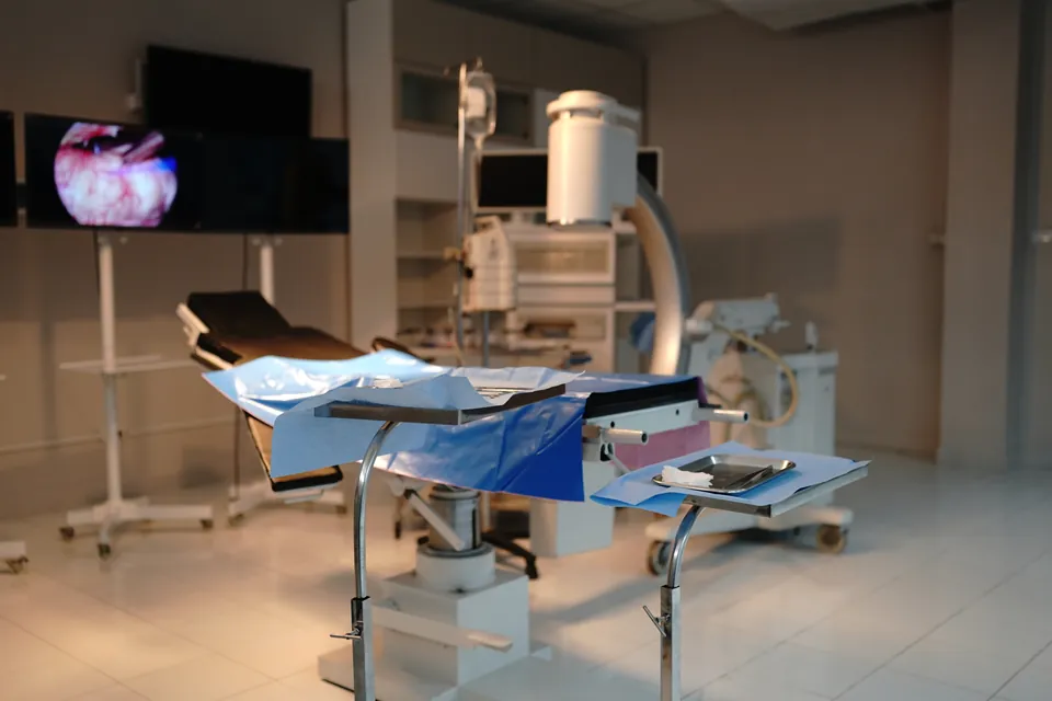
What is operative hysteroscopy, and how is it used for polyps, a septum or adhesions? General information on anaesthesia, risks, recovery and follow-up.
Operative hysteroscopy is a minimally invasive operation in which a problem inside the uterine cavity is seen with a thin camera called a hysteroscope and treated with small instruments passed through the same route. No incision is made in the abdomen; the instrument is advanced through the vagina and cervix. A polyp, a septum (tissue dividing the uterine cavity), adhesions, and some fibroids that extend into the cavity are conditions that can be addressed with this method. The information below provides a general framework and does not replace an individual treatment recommendation.
What is operative hysteroscopy?
Hysteroscopy allows the inside of the uterus to be seen directly. In operative hysteroscopy, in addition to viewing, an instrument intended for treatment is used; the problematic tissue is cut, shaved, or removed. The type of instrument and energy source used vary according to the procedure and the surgeon's preference.
How does operative hysteroscopy differ from diagnostic hysteroscopy?
The diagnostic procedure aims to view the uterine cavity and can mostly be performed with a thin instrument in an office setting. The operative procedure, because it involves an intervention, usually requires more preparation and can take more time. The office or operating theatre setting is chosen according to the scope of the procedure; more extensive procedures are usually planned under anaesthesia in the operating theatre. When a problem is seen during a diagnostic examination, when and in which setting the treatment will be carried out is discussed with the doctor.
When is operative hysteroscopy considered?
The situations below are examples that can be addressed with operative hysteroscopy. Which procedure is performed is determined by the condition itself, its location, and the person's goal.
How are polyps inside the uterus treated?
Polyps extending from the endometrium into the cavity can be seen and cut out. The removed tissue is sent for pathology. Whether the polyp is causing symptoms, and factors such as pregnancy plans and age, determine whether monitoring or treatment is the right question.
How is a uterine septum treated?
In septum resection, a congenital band of tissue in the uterine cavity is seen with the hysteroscope and cut, correcting the shape of the cavity. This is not a diagnosis but a procedure option. Not every septum requires surgery, and the decision is individual. Sources do not offer a single, unified recommendation on the contribution of resection to pregnancy outcomes.
How are intrauterine adhesions treated?
Adhesions can develop between the uterine walls after a previous procedure, infection, or surgery. Hysteroscopic adhesiolysis is the separation of these bands under direct vision. Surgery for adhesions is usually performed using this method; evidence for additional measures aimed at preventing the adhesions from re-forming is limited.
Can fibroids extending into the uterine cavity be removed this way?
Only the types of fibroid that extend into the uterine cavity can be removed by hysteroscopy; other types of fibroid require different methods.
What assessment and preparation take place before surgery?
Before the operation, ultrasound, diagnostic hysteroscopy if needed, or other imaging is used to determine the location and size of the problem. Regularly used medicines, allergies, bleeding disorders, and the possibility of pregnancy are reported to the doctor. In a person with heavy bleeding, anaemia is also assessed. Instructions such as fasting, stopping medicines, and the timing of the operation are given by the doctor; timing can vary according to the type of problem and the menstrual cycle.
What happens with anaesthesia and in the short term after surgery?
You discuss with the anaesthesia team which option is suitable among general or regional anaesthesia or sedation. After the operation, a period of observation is carried out; when you can go home depends on the scope of the procedure and your general condition. The surgical team monitors how much of the fluid used to keep the uterine cavity open has passed into the body. The operation is performed by Assoc. Prof. Dr. Nurettin Türktekin at Memorial Şişli Hospital.
What are the risks and complications of operative hysteroscopy?
Complications can also occur with minimally invasive operations of this kind. The main possibilities are:
- Perforation of the uterine wall (reported more often in the operative procedure than in the diagnostic procedure)
- Bleeding during or after the operation
- Infection
- Fluid overload related to fluid passing into the body; this increases when the procedure is prolonged and the blood vessels of the uterine muscle are reached
- Development of new adhesions inside the uterus
- Problems related to anaesthesia
- The problem not being fully resolved in the first session, requiring a second procedure in this situation
Which of these risks is more significant for you varies according to the procedure to be performed; you can raise your questions with the doctor at the pre-operative consultation.
What can you expect during recovery and follow-up?
Light bleeding or spotting and mild cramping in the days following the procedure are expected and decrease over time. Heavy bleeding or severe pain is not usual. The time to return to daily activities, work, sexual intercourse, and sport varies according to the scope of the procedure. The removed tissue is sent for pathology, and the result is shared at the follow-up appointment. After an adhesion or septum procedure, medicine or a follow-up hysteroscopy aimed at reducing recurrence may be planned. The suitable time to try for pregnancy is communicated by the doctor according to the scope of the procedure.
Frequently Asked Questions
Are a polyp and a fibroid removed in the same session?
If there is more than one finding inside the uterus, whether these are addressed in the same operation depends on the duration of the procedure and monitoring of the fluid medium. A larger or deeper fibroid may not be fully removed in a single session; whether a second session is needed is discussed at the pre-operative consultation.
What should I do if my period becomes lighter after an adhesion or septum procedure?
A marked decrease or delay in periods is a finding that needs to be assessed at the follow-up appointment, depending on the scope of the procedure. Report this to your doctor; they will decide whether a follow-up hysteroscopy is needed.
What should I ask before trying for pregnancy?
You can ask the doctor about the scope of the procedure, the condition of the uterine wall, and whether follow-up imaging is needed. The doctor will tell you the suitable time to try, according to the scope of the procedure.
Does operative hysteroscopy increase IVF success?
Resolving a problem inside the uterus can be part of the treatment plan for some people; however, the procedure has not been shown to generally increase IVF success. In selected situations, the benefit is assessed according to the finding.
Is operative hysteroscopy open surgery?
No. No abdominal incision is made; the instrument passes through the vagina and cervix, and the procedure uses a hysteroscope and small instruments passed along the same route.
Can every type of fibroid be removed by hysteroscopy?
No. Only fibroids extending into the uterine cavity can be removed this way; other types need other methods.
Does septum surgery guarantee a chance of pregnancy?
No. There is no clear, consistent recommendation on how much septum incision contributes to pregnancy outcomes; not every septum needs surgery either.
Which symptoms are expected after surgery, and which are unusual?
Light bleeding or spotting and mild cramps are expected in the days afterwards and decrease over time. Heavy bleeding or severe pain is unusual.
Why is the fluid used during surgery monitored?
The surgical team monitors how much of the fluid used to keep the uterine cavity open enters the body; fluid overload risk increases as the procedure lengthens and vessels in the uterine muscle are reached.
Is removed tissue sent for pathology?
Yes. Removed tissue is sent for pathology and the result is shared with you at follow-up.
Can adhesions recur after adhesion or septum surgery?
Yes, this is a known risk. Medicines or follow-up hysteroscopy may be planned to reduce recurrence, but evidence about the effect of additional measures is limited.
Who performs operative hysteroscopy, and where?
The operation is performed by Assoc. Prof. Dr. Nurettin Türktekin at Memorial Şişli Hospital.
Get Your Free Consultation
Fill out the form below and our team will contact you

