


What is sonohysterography (SIS), what does it show and how is it done? Indications, preparation, pain, risks and how it differs from HSG and hysteroscopy.
Sonohysterography (SIS) is an imaging method in which sterile saline (physiological serum) is given into your uterine cavity while a transvaginal ultrasound is performed, to assess the inner surface of your uterus and findings that affect the cavity. The saline separates the walls of the uterus from one another and allows the structures in the cavity to be seen more clearly on ultrasound. The information below offers a general framework and does not replace a diagnosis or treatment recommendation specific to you.
What is sonohysterography (SIS)?
On a routine transvaginal ultrasound, the uterine cavity looks like a closed slit. In SIS, saline is given into the cavity through a thin catheter; the cavity widens with this fluid, and bulges on the inner surface or changes in shape in the walls become visible. No X-ray is used and no contrast material is given. In this respect SIS is a form of ultrasound done with saline.
In which situations is SIS requested?
SIS may be requested if you are having difficulty conceiving, to assess your uterine cavity, and to investigate abnormal uterine bleeding. When a polyp, a fibroid extending into the uterine cavity, adhesions or a septum is suspected on ultrasound, clarifying this impression is also a reason. In the assessment of unexplained infertility, current guidelines recommend ultrasound (three-dimensional if possible) as the first method for the uterus, and state that no additional assessment is needed if the cavity looks normal on ultrasound. SIS is a method your doctor turns to, according to you, in situations not clarified by ultrasound.
How do you prepare for SIS, and how is the timing determined?
Your SIS appointment is mostly given for the days after your menstrual bleeding has ended; if you come on a day with bleeding, your doctor may postpone the procedure. If there is a possibility that you are pregnant or you have signs of an active pelvic infection, the procedure is postponed. During preparation you need to tell your doctor about the possibility of pregnancy, the medicines you use and your allergies; in particular, mention a latex allergy before the procedure, because part of the material used may contain latex. Your doctor decides whether a painkiller is needed and the other preparations.
What happens during and after the procedure?
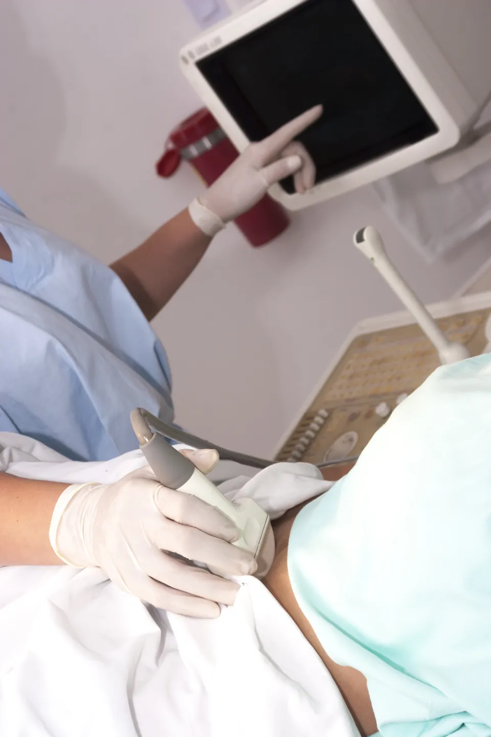
You lie in the gynaecological examination position and a speculum is used during the examination. While saline is given through a thin catheter passed through your cervix, your uterine cavity is watched live with the ultrasound probe in the vagina; as the cavity fills with fluid, the walls and the bulges inside are scanned on the screen. No X-ray image is taken, so the focus is the ultrasound sections on the screen. You may feel a cramp similar to period pain; the pain varies from person to person. The duration varies with the findings seen and with the person.
On leaving, mild cramp and light spotting are common.
What does SIS show, and what does it not show?
SIS can show findings in your uterine cavity such as a polyp, a fibroid extending into the cavity, adhesions and a septum. SIS is counted among the methods used to detect lesions inside the uterus. Even so, your doctor interprets what the imaging finding is, and the distinction between a polyp, a fibroid or adhesions may not be clear in the image. Very fine adhesions may be missed. SIS has a high predictive value in detecting findings inside the uterus; even so, SIS does not make a definitive diagnosis on its own, and for lesions in the uterine cavity the definitive diagnosis and treatment method is hysteroscopy.
SIS is not the main method for showing tubal patency; other methods are used for this purpose. SIS is not done for the purpose of treatment, and it has not been shown to increase the likelihood of pregnancy.
What is the difference between SIS, HSG and hysteroscopy?
| SIS | HSG | Hysteroscopy | |
|---|---|---|---|
| Imaging | Ultrasound, saline | X-ray, contrast material | Direct viewing with a camera |
| Main purpose | Uterine cavity | Tubal patency and shape of the cavity | Inner surface of the uterus |
| Tubal patency | Gives limited information | Shows it | Not its routine purpose |
| Radiation | None | Yes | None |
| Invasiveness and pain | A catheter is needed; cramp may occur | A catheter is needed; cramp may occur | May be more invasive; pain and need for anaesthesia vary |
| Treatment | Its purpose is diagnosis | Its purpose is diagnosis; a therapeutic effect has not been proven | May be possible in the same procedure |
The choice is made according to your complaint, your history, your allergies, your access and your doctor's preference. None automatically takes the place of another.
What are the risks of SIS, and in whom is it not done?
SIS is generally a well-tolerated procedure, but it cannot be said to have no risks. The main problems are pain and cramp, a temporary feeling of dizziness, light spotting and, rarely, pelvic infection; if you have a history of pelvic infection, your doctor may plan an additional precaution. If the dizziness does not pass once you leave the room, gets worse or leads to fainting, it counts as urgent. A latex allergy is an important point. The procedure is not done if there is a possibility that you are pregnant or you have an active pelvic infection. A normal SIS result does not mean you will become pregnant.
How is the SIS result interpreted, and what is the next step?
A normal result shows that no structural problem is seen in your uterine cavity; the remaining assessment turns to the other possible causes of your difficulty in conceiving. If there is an abnormal finding, your doctor decides, according to you, whether it needs to be confirmed by hysteroscopy or whether treatment needs to be planned.
Frequently Asked Questions
Is an X-ray taken during SIS?
No. No X-ray is used and no contrast material is given in SIS; SIS is a form of ultrasound done with saline.
Is SIS done during pregnancy?
No. The procedure is not done if there is a possibility that you are pregnant or you have an active pelvic infection.
Can SIS be done if I have a latex allergy?
You need to tell your doctor this information beforehand; part of the material used may contain latex. With this information your doctor can re-plan the preparation and the procedure.
Does SIS show whether my tubes are open?
No. SIS is not the main method for showing tubal patency; other methods are used for this purpose.
Is SIS a treatment, and does it increase my chance of pregnancy?
No. SIS is not done for the purpose of treatment, and it has not been shown to increase the likelihood of pregnancy.
Does a finding seen on SIS mean a definitive diagnosis?
No. Although SIS has a high predictive value, it does not make a definitive diagnosis on its own. For lesions in the uterine cavity, the definitive diagnosis and treatment method is hysteroscopy.
Does a normal SIS result mean I will become pregnant?
No. A normal result shows that no structural problem is seen in your uterine cavity; the remaining assessment turns to the other possible causes. A normal result does not mean you will become pregnant.
Which symptoms are usual after SIS?
On leaving, mild cramp and light spotting are common.
Is it normal to feel dizzy during SIS?
A temporary feeling of dizziness may be seen. However, if it does not pass once you leave the room, gets worse or leads to fainting, it counts as urgent.
How is the choice made between SIS and HSG?
What is decisive is the question to which an answer is sought: a finding in the uterine cavity, or whether the tubes are open? In addition, your allergy history and the possibility of pregnancy influence your doctor's decision. Share this information clearly at the appointment.
What can be done to reduce pain during SIS?
Whether a painkiller is needed before the procedure is discussed with your doctor. Relaxation and deep breathing techniques may also help; if the pain is increasing, it is enough to tell the person performing the procedure.
What happens if the SIS result is abnormal?
An abnormal finding does not in itself require surgery. Your doctor assesses the finding together with your complaint and your other results. At the results consultation it is enough to ask: "What does this finding mean, and is monitoring or an additional investigation needed?"
When do I learn the SIS result?
Your doctor can share the findings on the screen with you during or straight after the procedure. The timing of the written report and the follow-up consultation varies from centre to centre; ask about this before the procedure.
Get Your Free Consultation
Fill out the form below and our team will contact you

