


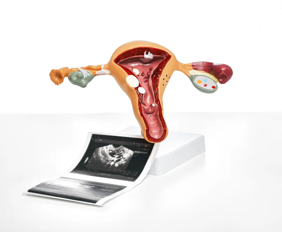
A septate uterus has a congenital band of tissue dividing the cavity. Symptoms, diagnosis, the link with pregnancy and treatment options explained.
A septate uterus contains a congenital tissue band, or septum, partly or completely dividing the uterine cavity. Many women have no symptoms, and effects on pregnancy outcomes vary. The outside of the uterus appears normal; the difference is inside. This information is general, not individual diagnostic or treatment advice.
What is a septate uterus?
The uterus forms during embryonic development from the fusion of two separate ducts. After this fusion, the wall between them should disappear completely. If part or all of this wall remains, a septate uterus results. The length and thickness of the septum vary from person to person, so a septate uterus does not have a single uniform appearance. Under the current classification system, the septate uterus is class U2, defined as a uterus with a normal outer contour and an internal indentation at the midline of the fundus.
Is a septate uterus the same as a bicornuate uterus?
No. Bicornuate and didelphys types also have a different external shape; a septate uterus has a normal outer contour and a divided cavity. The distinction matters because pregnancy outcomes and treatment differ. Imaging and further investigations if needed establish the type.
Why does a septate uterus develop?
A septate uterus is congenital, arising during the development of the uterus; it is not acquired later in life. Unlike adhesions that develop after surgery or a procedure, the septum has been present since birth.
What symptoms does it cause, and how is it discovered?
A septate uterus most often causes no symptoms. The menstrual pattern is usually preserved. The diagnosis is often made incidentally, either during an assessment for difficulty conceiving or recurrent pregnancy loss, or during imaging carried out for another reason.
How is a septate uterus diagnosed?
A single method may not always be sufficient to assess the shape of the uterus; the doctor chooses according to the symptom and the initial findings.
- Three-dimensional transvaginal ultrasound: current medical guidance identifies this as the first-line diagnostic tool for assessing uterine shape; it can be performed with or without saline.
- Hysterosalpingogram (HSG): shows the shape of the uterine cavity; because it does not show the outer contour, it can be limited on its own for distinguishing a septate from a bicornuate uterus.
- MRI: can provide additional information when ultrasound is insufficient or the distinction cannot be made clearly.
- Hysteroscopy: allows direct visualisation from inside the uterine cavity. More than one investigation is sometimes needed.
Diagnostic criteria differ between classification systems; the same uterus may be classified differently under different systems. For this reason, which criterion a report's description is based on also matters.
Can a septate uterus affect conception or pregnancy loss risk?
The effect of a septate uterus on pregnancy outcomes varies from person to person. The septum is thought to possibly increase the risk of pregnancy loss and preterm birth; however, the evidence on this is conflicting, so the level of risk cannot be stated and the same outcome cannot be predicted for every woman. This information is for general guidance; a person planning pregnancy assesses their own situation together with their doctor.
Does everyone with a septate uterus need septum incision surgery?
No. Septum incision uses hysteroscopy to divide the septum and restore the cavity's normal shape. Sources differ in whom they recommend it for. One current medical view offers it as an option through shared decision-making for a septum with recurrent miscarriage. In infertility, it considers incision reasonable but cannot give a definitive recommendation because improved live birth rates are uncertain. A multicentre randomised trial found no improvement in reproductive outcomes after incision. Another current review of recurrent pregnancy loss reports reduced loss rates in low-quality studies but no demonstrated effect on clinical pregnancy or live birth. Because the single small randomised trial showed no benefit, the recommendation is conditional and larger studies are needed. Sources therefore differ in scope and emphasis; there is no one-directional recommendation. Decide with your doctor according to history, age, pregnancy goals and previous outcomes.
What happens after hysteroscopic septum incision?
Current medical guidance regards incision as low risk, but known procedural risks include uterine perforation, bleeding, infection, adhesions and fluid overload. Your doctor determines review timing and when to try for pregnancy.
How is pregnancy monitored with a septate uterus?
In someone with a septate uterus, pregnancy is managed with close monitoring by a doctor who is aware of the risks. If bleeding, abdominal pain, waters breaking or a change in discharge occurs during pregnancy, you must seek care at a healthcare facility without delay.
Frequently Asked Questions
Is a septate uterus often found incidentally?
Yes. As it usually causes no symptoms, it is often discovered during assessment of difficulty conceiving or recurrent pregnancy loss, or imaging for another reason.
Does a septate uterus disturb periods?
No. Period patterns are usually preserved.
Is one imaging method sufficient for diagnosis?
Not always. Three-dimensional ultrasound is the first-line tool, but HSG, MRI or hysteroscopy may be needed to assess the external contour or clarify uncertain distinctions. Several examinations may be assessed together.
Does a septate uterus definitely cause pregnancy loss?
No. It may increase miscarriage and preterm birth risk, but evidence conflicts and effects vary. The same outcome cannot be predicted for every woman.
Is hysteroscopic incision recommended for everyone with a septum?
No. Not every septum needs surgery. Decide with your doctor according to history, age, pregnancy goals and previous outcomes.
Does incision definitely increase live birth rates?
Sources do not agree. Some studies report reduced pregnancy loss, but an effect on clinical pregnancy or live birth is unproven; a multicentre randomised trial also found no improvement in reproductive outcomes. A one-directional recommendation cannot therefore be made.
Is septum incision a risky operation?
It is considered low risk, but known risks include uterine perforation, bleeding, infection, adhesions and fluid overload.
Can I try for pregnancy immediately afterwards?
Your doctor determines review timing and when to try; this varies individually.
Is septum resection performed for every uterine shape abnormality?
No. According to a current review of recurrent pregnancy loss, in people with recurrent pregnancy loss, metroplasty or reconstruction is not recommended for a bicorporeal uterus with a normal cervix (formerly called bicornuate) or a hemi-uterus; for a bicorporeal uterus with two cervices (formerly called didelphys), the evidence is insufficient. It should be remembered that the septum is a separate shape abnormality from these conditions; which type is present is determined by imaging.
I had a miscarriage - is a septate uterus the cause?
The septum alone may not be the explanation. In recurrent pregnancy loss, the shape of the uterus is only one of the causes assessed; other factors are also investigated separately. Finding a septum does not mean that the septum was the cause of the miscarriage.
Are a septate uterus and a vaginal septum the same thing?
No. A septate uterus refers to tissue dividing the uterine cavity, while a vaginal septum refers to tissue dividing the vagina. The two can occur together, so the vagina and cervix are also assessed during examination and imaging.
Get Your Free Consultation
Fill out the form below and our team will contact you

