


What is embryo biopsy, at which stages is it done, and what happens in trophectoderm biopsy? Possible effects on the embryo, limits and doctor questions.
Embryo biopsy is the taking of a small number of cells from an embryo obtained through IVF for genetic examination (PGT). Today it is most often done at the blastocyst stage, from the outer layer of the embryo (the trophectoderm). It does not make a diagnosis on its own; the sample taken is examined with a separate test. Because it is an intervention on the embryo, it has risks such as the possibility of harming the embryo, and the sample taken may not always reflect the whole embryo. Your doctor and embryologist assess whether it will be considered for you.
Biopsies taken from the cervix or from the lining of the uterus are different procedures; embryo biopsy refers only to sampling done on the embryo in the laboratory.
Does embryo biopsy make a diagnosis on its own?
The function of the biopsy is to provide a sample containing DNA for genetic testing. After the sample is taken it is examined in the genetic laboratory, and the diagnostic or screening result comes from this separate test.
For which tests can the biopsy sample be used?
The sample taken can be used in PGT-A, which screens chromosome number, in PGT-M, which investigates single-gene diseases, and in PGT-SR, which is aimed at structural chromosome changes. The purpose and the state of evidence of these tests are assessed separately.
Can a sample be taken from the polar body or at the cleavage stage?
According to the current recommendations on this subject, a sample can be taken from the polar bodies of the egg or from a single cell of the embryo at the cleavage stage. Cleavage-stage biopsy was widely applied for a time, but its clinical use has declined.
Why is blastocyst biopsy described as the commonly used technique?
The blastocyst is a more advanced stage of the embryo. According to the current assessment on this subject, taking a sample from the outer layer at this stage is at present the most widely used technique and provides more cells than the cleavage stage. This does not mean that the method is superior to the others; this assessment only describes present practice.
How is the procedure performed?
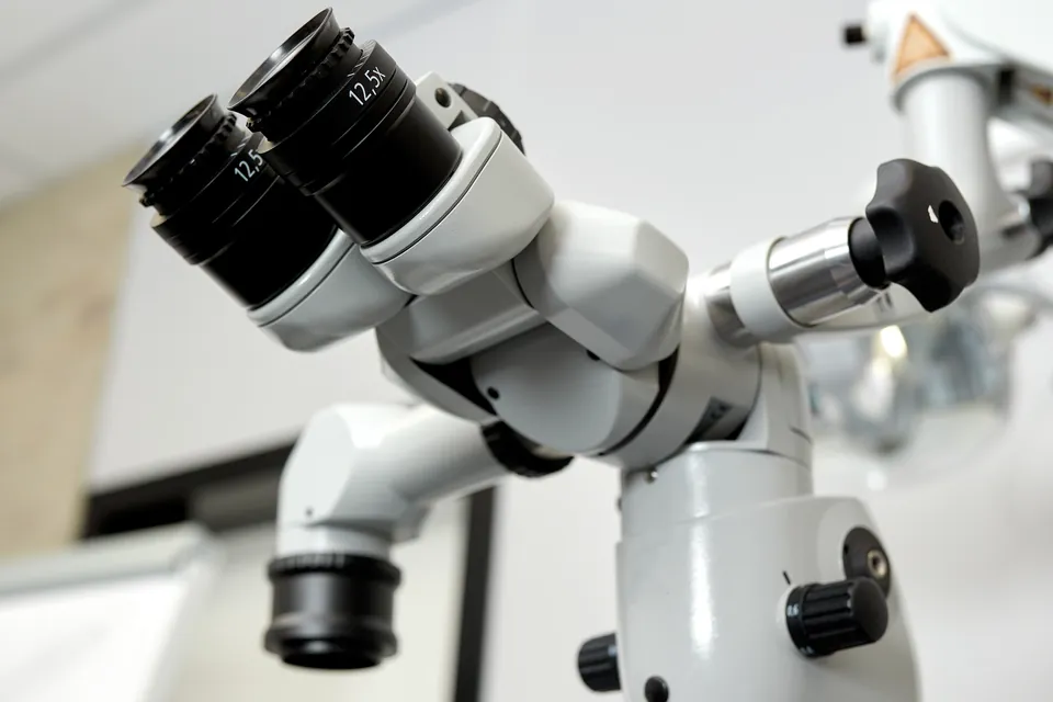
The procedure is performed on the embryo in the laboratory, under the microscope, by embryology specialists. The shell that surrounds the embryo (zona pellucida) is opened; the current recommendations state that a laser is most often used for this purpose today. Then a small number of cells are taken from the outer layer and transferred into a separate tube to be sent to the genetic laboratory. The same recommendations advise that in blastocyst biopsy the inner cell mass be protected and as little intervention as possible be applied. The number of cells, the duration of the procedure and technical details vary with the laboratory.
What happens after the biopsy?
Because the result is not available immediately, biopsied embryos are generally frozen. The current recommendations on this subject state that freezing is mostly necessary in situations where the test result must be waited for. When the result comes, which embryo will be transferred, and when, is decided together with your doctor. The freezing and thawing process has its own limits; this is a separate heading.
Can the biopsy harm the embryo?
According to the current assessments on this subject, taking cells can harm the embryo and hinder its development. The same assessment states that the biopsy is invasive and can damage cells and DNA. According to another current view, the effect of trophectoderm biopsy is not well understood. For this reason it is expected that these possibilities will be addressed at the consent consultation.
Does the sample taken reflect your whole embryo?
According to the current mosaicism recommendations on this subject, a finding detected in the outer layer may not show the state of the embryo as a whole or its developmental potential. The interpretation of this situation should be dealt with separately in genetic counselling.
Is every embryo suitable for biopsy?
Not every embryo may be suitable for biopsy. The current assessment on this subject reports that embryos may be lost because they are not suitable for biopsy. Your doctor and embryologist decide which embryos are suitable.
Is embryo biopsy the same as assisted hatching?
No. Assisted hatching aims at thinning or opening the shell in order to help the embryo attach to the uterus. The purpose of biopsy, on the other hand, is to take a cell sample for genetic examination. In the current recommendations on this subject, opening the shell is mentioned as a step in the procedure of taking cells; but this does not turn the biopsy into a hatching practice. It is sensible to clarify with your doctor and embryologist which one is planned.
What can you ask your doctor and embryologist?
- At which stage is the biopsy planned, and why?
- How many of my embryos are expected to be suitable for biopsy?
- What will happen to embryos that cannot be biopsied?
- How will the embryos be stored after the biopsy?
- How is the possibility of harming the embryo expressed in the consent text?
Frequently Asked Questions
Does the biopsy carry a procedure risk for me?
According to the current assessment on this subject, there is no known additional risk for the person receiving PGT-A treatment. Because the procedure is performed on the embryo in the laboratory, the risks are at the level of the embryo. The risks belonging to stages of the IVF cycle such as ovarian stimulation and egg collection apply independently of the biopsy.
Can the biopsy be repeated?
According to the current assessment on this subject, re-biopsy is done only when the first procedure has failed, was incomplete or was inconclusive, and its effect on development is still being studied. Re-biopsy is not recommended in order to confirm a mosaic result; according to the current mosaicism recommendations on this subject, there is no evidence that this has diagnostic value.
Is there data on children born after biopsy?
According to the current assessment on this subject, studies following children born from embryos biopsied at the blastocyst stage have not revealed an adverse picture regarding pregnancy and birth. However, a small increase in a degree of delay in growth inside the uterus may require separate investigation. The same assessment also states that there is uncertainty about the long-term effects of IVF and PGT.
Are embryo biopsy and genetic testing the same thing?
No. Biopsy is taking a cell sample for genetic testing. The diagnostic or screening result comes from the separate test in which the sample is examined in the genetic laboratory.
Is it the same procedure as a biopsy of the lining of the uterus?
No. Embryo biopsy is done on the embryo in the laboratory. Biopsies of the cervix and of the lining of the uterus are different procedures.
Does blastocyst biopsy being the most common mean it is superior?
No. This statement describes current practice. Providing more cells than the cleavage stage does not mean that it is superior to the other methods.
Is the protection of the embryo's inner cell mass taken into account when the sample is taken?
Yes. The current recommendations advise that in blastocyst biopsy the inner cell mass be protected and as little intervention as possible be made.
Is every one of your embryos suitable for biopsy?
Not every embryo may be suitable. Your doctor and embryologist assess suitability; you can ask beforehand how embryos not suitable for biopsy will be handled.
Does a mosaic finding represent the whole embryo?
It may not always represent it. A finding in the outer layer may not show the state of the embryo as a whole or its developmental potential; the interpretation is dealt with in genetic counselling.
Can embryos be frozen while waiting for the result?
Yes. Because the result is not available immediately, biopsied embryos are generally frozen. The freezing and thawing process also has its own limits.
Do biopsy and assisted hatching serve the same purpose?
No. Biopsy provides a cell sample for genetic examination. Assisted hatching aims at thinning or opening the shell that surrounds the embryo in order to help attachment.
Get Your Free Consultation
Fill out the form below and our team will contact you

